Every view is found the same way: choose the probe, position the patient, place the probe with its marker the right way, sweep and tilt until the view appears, then name each structure by how bright it looks. Each view below gives the probe steps and what a good view shows. The vessel and musculoskeletal views are tagged later: the objectives name them, but no source covers them, so only the objectives are given.
Pick a region below to jump to it.
Choose the probe → Position the patient → Place the probe, marker the right way → Sweep and tilt → Name each structure
The probe marker points to the patient's head (or to their right in a cross-section). Fluid is black, solid organs are grey, bone and the diaphragm are bright white.
General approach
Every ultrasound
The general approach
Say it first (TOSCE): "Hello, I'm ___, a medical student. Can I check your name and date of birth? I'd like to do a quick bedside ultrasound of your [heart / abdomen]. I'll put some cool gel on your skin and press gently with the probe. It doesn't hurt and uses no radiation. Is that alright?" Then wash your hands, lie the patient flat on their back (supine), and uncover only the area you are scanning.
Choose the probe: the curved (curvilinear) probe, or the small flat-faced phased array probe, for the heart and abdomen: they use lower-frequency sound that reaches deeper
Probe marker: the ridge or dot on one side of the probe; it matches a dot on the screen
for a lengthwise view (sagittal front-to-back slice, or coronal side-on slice) the marker points to the patient's head
for a cross-section (transverse / axial) it points to the patient's right
Reading the screen: the top of the screen is the skin under the probe (near field); the bottom is deepest (far field)
Words for brightness (echogenicity)
anechoic = black: fluid (blood, bile, urine)
hypoechoic = darker grey than the tissue around it
isoechoic = the same grey as the tissue around it (liver, spleen, kidney cortex)
hyperechoic = bright white: bone, the diaphragm, fat around the kidney
Acoustic window: sound travels well through solid organs and fluid, badly through air and bone. So you look through the liver (to the heart, the kidney), or through a full bladder (to the pelvis)
Heart: the subxiphoid view
PoM 1 · POCUS session · 1 views
Before you scan
The view
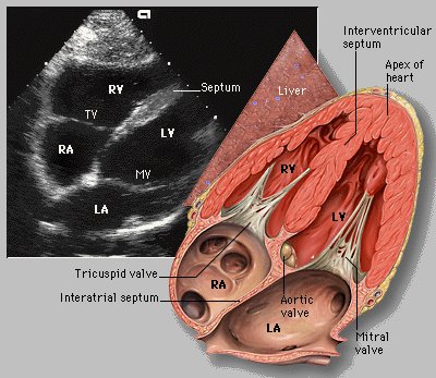
The probe sits just below the breastbone (under the xiphoid process) and looks up through the liver at the heart; the liver is the window, lying directly under the probe
Name the structures: right ventricle, left ventricle, right atrium, left atrium, interventricular septum (the wall between the ventricles), pericardium (the sac around the heart)
Show the cardiac cycle: systole (the ventricles squeeze and get smaller) and diastole (they relax and fill)
Is the heart moving, and is there fluid around it?
Subxiphoid (heart)
All four chambers, the septum and the pericardium, beating
Picture
Steps
Patient supine; curved or phased array probe
Start with the probe flat on the umbilicus and slide it up to the xiphoid
Look up through the liver at the heart
Can't see the heart? Inspiratory hold (a deep breath held pushes the diaphragm down and brings the heart closer) · bend the knees (relaxes the abdominal muscles)
A good view
Good view = liver nearest the probe, then the heart, with right and left ventricles, right and left atria and the septum visible, the pericardium around them, moving through systole and diastole
Upper abdomen
PoM 1 · POCUS session · 3 views
Before you scan
The view
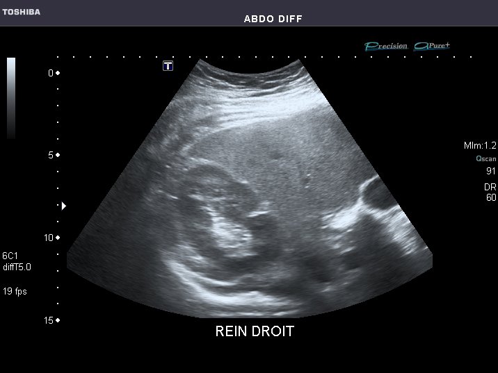
Right upper quadrant (RUQ): liver, right kidney, diaphragm
liver = isoechoic (even grey)
kidney = a "double density": grey outer cortex, bright white centre (the renal sinus: collecting system with the calyces and renal pelvis, fat and the main blood vessels)
the slit between liver and kidney is the hepatorenal space (Morison's pouch)
diaphragm = a bright white curved line above the liver
the spine shows deep in the far field as a bright "railroad track"
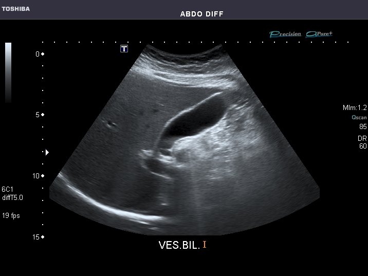
Gallbladder: the gallbladder and the portal triad (portal vein, hepatic artery, common bile duct)
gallbladder = anechoic (black, full of bile)
the exclamation-point sign: the gallbladder is the line, the portal triad is the dot (shaped like a Mickey Mouse head), and the main lobar fissure (a thin bright line in the liver) joins them
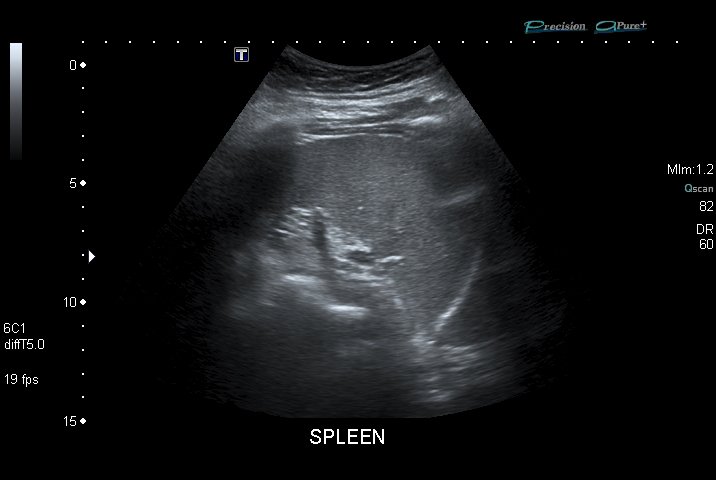
Left upper quadrant (LUQ): spleen, left kidney, diaphragm
spleen = isoechoic · left kidney = double density · diaphragm = bright white · the lung sits above the diaphragm
Liver, kidney and the space between them
Right upper quadrant
Liver, right kidney, diaphragm
Picture
Steps
Patient supine; curved probe
Probe on the right side, mid-axillary line, level with the xiphoid, in a coronal (side-on) plane
Marker to the patient's head
Slide toward the feet or the head until the kidney appears
A good view
Good view = liver above, kidney below, Morison's pouch between them, the diaphragm as a bright line, the spine deep
Gallbladder
Gallbladder and portal triad
Picture
Steps
Probe under the right costal margin (edge of the ribs), midclavicular line
Sagittal plane, marker to the patient's head
Fan slowly until a black, bag-shaped structure appears
A good view
Good view = the exclamation point: black gallbladder (line), main lobar fissure, portal triad (dot)
Left upper quadrant
Spleen, left kidney, diaphragm
Picture
Steps
Same set-up as the RUQ, on the left side, marker to the head
Slide toward the feet or the head until the kidney appears
A good view
Good view = spleen, left kidney below it, the diaphragm as a bright line, lung above
Pelvis
PoM 1 · POCUS session · 2 views
Before you scan
The view
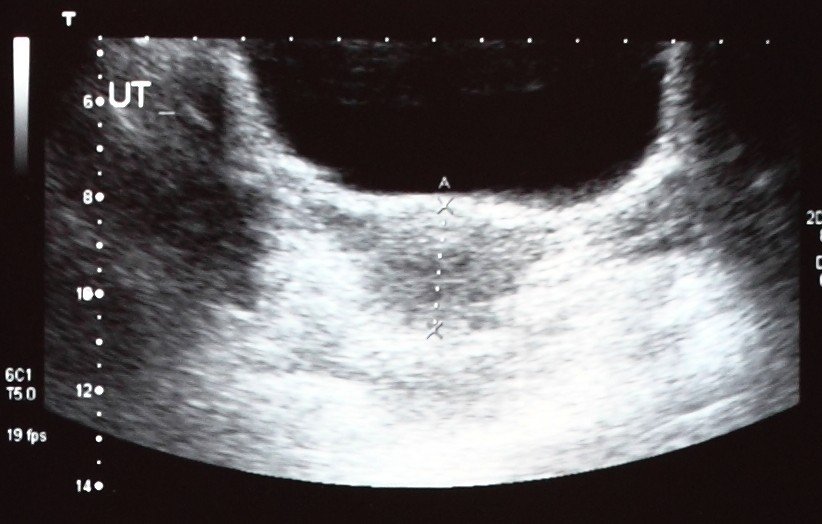
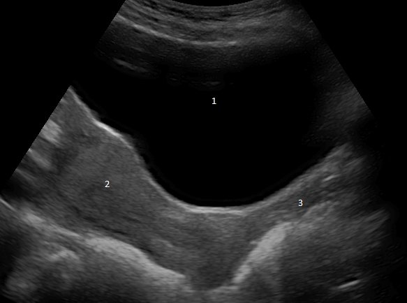
Name the structures: bladder, uterus or prostate, rectum
The bladder must be full: urine is the acoustic window into the pelvis
Male: bladder (anechoic) → prostate below it → rectum below that; note the rectovesical space (between bladder and rectum)
Female: bladder (anechoic) → uterus (isoechoic) in the far field, with a bright line in its centre (the endometrium, the uterine lining) → rectum below; note the vesicouterine space (between bladder and uterus) and the rectouterine pouch (pouch of Douglas) behind the uterus
in the sagittal view you also see the vagina as a darker (hypoechoic) band behind the bladder
Bladder and what lies behind it
Pelvis, transverse
Bladder, uterus or prostate, rectum
Picture
Steps
Patient supine with a full bladder; curved probe
Probe across the lower abdomen (transverse), marker to the patient's right
Slide down to the pubic symphysis (pubic bone), then tilt the probe down into the pelvis
A good view
Good view = black bladder at the top, uterus or prostate behind it, rectum deepest
Pelvis, sagittal
The same structures, lengthwise
Picture
Steps
From the transverse view, rotate the probe to sagittal, marker to the head
A good view
Good view = bladder, uterus (with endometrium) or prostate, vagina, rectum in a line
Vessels
T2C · POCUS
Before you scan
The view
No source covers these views in detail: only the objectives are given here; learn the probe positions in session
Abdominal aorta view: identify the abdominal aorta and the inferior vena cava (IVC)
Carotid sheath view (the bundle in the side of the neck): identify the carotid artery and the internal jugular vein (IJV)
Valsalva (bearing down against a closed throat): show its effect on the internal jugular vein
Compression with the probe: show its effect on the internal jugular vein and the carotid artery
Musculoskeletal structures
T2C · POCUS
Before you scan
The view
Not covered by the sources used here: only the objectives are given here
Achilles tendon: show the view and identify the linear (fibre-like) pattern of its collagen fibres
Arm: show the view and identify bone, muscle and nerve
Posterior shoulder joint: show the view and identify the humeral head, the glenoid and the infraspinatus tendon
Video credits
Ultrasound screenshots are reused under their open licences, credited below. Each view also has a probe-position video from Dr. Sam's Imaging Library, played from YouTube only when you tap it. Practise the probe movements on a classmate in the session.
Image credits (6)
Images used for non-commercial teaching under their own licences (Creative Commons or public domain; textbook figures from student notes remain their publishers' copyright). Some were cropped to hide faces or handwriting.
Subxiphoid (heart): Patrick J. Lynch, medical illustrator, and C. Carl Jaffe, MD, CC BY 2.5, source
Right upper quadrant: Ptrump16, CC BY-SA 4.0, source